Córtex retrosplenial
Córtex retrosplenial
| |
|---|---|
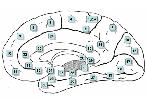 Superfície medial do cérebro com as áreas de Brodmann numeradas
| |
| Identificadores | |
| Latim | regio retrosplenialis |
O córtex retrosplenial (CRS) é uma área cortical do cérebro composta pelas áreas de Brodmann 29 [en] e 30 [en].[1] Trata-se de um córtex de associação secundária, estabelecendo conexões com numerosas outras regiões cerebrais. O nome da região faz referência à sua localização anatômica imediatamente posterior ao esplênio do corpo caloso em primatas, embora em roedores esteja situado mais próximo da superfície cerebral e apresente tamanho relativamente maior. Sua função ainda não é totalmente compreendida, mas sua proximidade com áreas visuais e também com o sistema espacial e de memória do hipocampo sugere que possa desempenhar um papel de mediação entre funções perceptivas e de memória,[2] particularmente no domínio espacial.[3] Contudo, sua contribuição exata para o processamento do espaço ou da memória tem sido difícil de determinar com precisão.[4]
Anatomia
Existe grande variação no tamanho dessa região entre diferentes espécies. Em humanos, ela corresponde a aproximadamente 0,3% de toda a superfície cortical, enquanto em coelhos representa pelo menos 10%[5] e, em ratos, estende-se por mais da metade do cérebro no sentido dorsoventral, constituindo uma das maiores regiões corticais.[2]
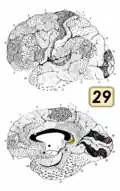
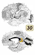
Com base em sua estrutura celular microscópica, o córtex retrosplenial é dividido em regiões disgranular (área 30) e granular (área 29).[1] Essas subdivisões foram posteriormente organizadas em quatro áreas: 29l, 29m, 30l e 30m.[6] O córtex apresenta projeções recíprocas densas com o córtex visual, com o pós-subículo (também conhecido como pré-subículo dorsal), com os núcleos talâmicos anteriores e com o hipocampo.[7]
Neurofisiologia
Estudos neurofisiológicos do córtex retrosplenial foram realizados principalmente em ratos. Trabalhos iniciais demonstraram que aproximadamente 8,5% dos neurônios dessa região são células de orientação da cabeça [en], enquanto outros neurônios apresentam correlação com parâmetros de movimento, como localização espacial e velocidade de deslocamento.[8][9] Pesquisas mais recentes indicam que a atividade neuronal do córtex retrosplenial reflete simultaneamente múltiplos parâmetros, incluindo o ambiente em que o animal se encontra,[10] sua posição espacial dentro dele,[11][10] a direção atual da cabeça e a velocidade de corrida,[10] bem como se o animal está realizando uma curva[11] ou planeja virar no futuro.[12] Muitas dessas características neurofisiológicas desenvolvem-se gradualmente à medida que o animal aprende a navegar em um ambiente,[12] o que é consistente com a hipótese de que o córtex retrosplenial participa do armazenamento de longo prazo da memória espacial.[13]
Função
Em humanos, estudos de imagem por ressonância magnética funcional implicam o córtex retrosplenial em uma ampla variedade de funções cognitivas, incluindo memória episódica, navegação, imaginação de eventos futuros e processamento de cenas de modo geral.[2][14] Estudos em roedores sugerem que a região é importante para utilizar pistas visuais do ambiente na execução dessas tarefas.[13][15][16][17] O córtex retrosplenial é especialmente sensível a marcos ambientais permanentes e estáveis[18][19] e também está envolvido em utilizá-los para realizar julgamentos espaciais.[20][21] Uma revisão publicada em 2023 sugeriu que sua função principal estaria relacionada à “navegação espacial e localização de objetos pessoalmente relevantes”.[6]
Também foi proposto que o córtex retrosplenial possa atuar na tradução entre informações espaciais egocêntricas (centradas no indivíduo) e alocêntricas (centradas no mundo), devido à sua posição anatômica intermediária entre o hipocampo (onde existem representações alocêntricas por meio das células de lugar) e o lobo parietal (que integra informações sensoriais egocêntricas).[13][22][23]
Competidores do Campeonato Mundial de Memória são capazes de realizar feitos excepcionais de memória e apresentam maior ativação no córtex retrosplenial em exames de imagem por ressonância magnética funcional em comparação com indivíduos comuns.[24] Acredita-se que isso se deva ao uso de uma estratégia de aprendizado espacial ou técnica mnemônica conhecida como palácio da memória.
A região também apresenta ritmicidade de ondas lentas do tipo ritmo teta[25] e, quando pessoas evocam memórias autobiográficas, ocorre interação na faixa teta entre o córtex retrosplenial e o lobo temporal medial.[26]
Patologia
O córtex retrosplenial é uma das diversas áreas cerebrais cuja lesão pode produzir tanto amnésia anterógrada quanto amnésia retrógrada.[27] Pessoas com lesões envolvendo essa região também podem apresentar uma forma de desorientação topográfica [en], na qual conseguem reconhecer e identificar marcos ambientais, mas são incapazes de utilizá-los para orientar-se no espaço.[2]
O córtex retrosplenial é uma das primeiras regiões a sofrer alterações patológicas na doença de Alzheimer e em sua fase prodrômica, o declínio cognitivo leve.[28][29][30] Existem ainda evidências experimentais indicando que a camada 5 do córtex retrosplenial pode estar envolvida na geração de estados dissociativos de consciência em mamíferos.[31]
Galeria
Referências
- ↑ a b Vogt, B. A. (1 de setembro de 1976). «Retrosplenial cortex in the rhesus monkey: a cytoarchitectonic and Golgi study». The Journal of Comparative Neurology. 169 (1): 63–97. ISSN 0021-9967. PMID 821976. doi:10.1002/cne.901690105
- ↑ a b c d Vann, Seralynne D.; Aggleton, John P.; Maguire, Eleanor A. (8 de outubro de 2009). «What does the retrosplenial cortex do?». Nature Reviews Neuroscience. 10 (11): 792–802. PMID 19812579. doi:10.1038/nrn2733
- ↑ Mitchell, Anna S.; Czajkowski, Rafal; Zhang, Ningyu; Jeffery, Kate; Nelson, Andrew J. D. (19 de março de 2018). «Retrosplenial cortex and its role in spatial cognition». Brain and Neuroscience Advances (em inglês). 2. PMC 6095108
 . PMID 30221204. doi:10.1177/2398212818757098
- ↑ Vann, Seralynne D.; Aggleton, John P.; Maguire, Eleanor A. (novembro de 2009). «What does the retrosplenial cortex do?»
 . Nature Reviews Neuroscience (em inglês). 10 (11): 792–802. ISSN 1471-003X. PMID 19812579. doi:10.1038/nrn2733
- ↑ notes, K. Brodmann; translated with editorial; Garey, an introduction by Laurence J. (2006). Brodmann's Localisation in the cerebral cortex the principles of comparative localisation in the cerebral cortex based on cytoarchitectonics 3rd ed. New York: Springer. ISBN 978-0-387-26919-1
- ↑ a b Vogt, Brent A.; Rosene, Douglas L. (2023). «Comparison of monkey and human retrosplenial neurocytology». Journal of Comparative Neurology. 531 (18): 2044–2061. ISSN 0021-9967. doi:10.1002/cne.25561
- ↑ Sugar, Jorgen; Witter, Menno P.; van Strien, Niels; Cappaert, Natalie (2011). «The Retrosplenial Cortex: Intrinsic Connectivity and Connections with the (Para)Hippocampal Region in the Rat. An Interactive Connectome». Frontiers in Neuroinformatics (em inglês). 5: 7. ISSN 1662-5196. PMC 3147162. PMID 21847380. doi:10.3389/fninf.2011.00007
- ↑ Chen, Longtang L.; Lin, Lie-Huey; Green, Edward J.; Barnes, Carol A.; McNaughton, Bruce L. (setembro de 1994). «Head-direction cells in the rat posterior cortex». Experimental Brain Research. 101 (1): 8–23. PMID 7843305. doi:10.1007/BF00243212
- ↑ Cho, J; Sharp, PE (fevereiro de 2001). «Head direction, place, and movement correlates for cells in the rat retrosplenial cortex.». Behavioral Neuroscience. 115 (1): 3–25. PMID 11256450. doi:10.1037/0735-7044.115.1.3
- ↑ a b c Miller, Adam M P; Serrichio, Anna C; Smith, David M (1 de maio de 2021). «Dual-Factor Representation of the Environmental Context in the Retrosplenial Cortex». Cerebral Cortex. 31 (5): 2720–2728. ISSN 1047-3211. PMC 8023839. PMID 33386396. doi:10.1093/cercor/bhaa386
- ↑ a b Alexander, Andrew S.; Nitz, Douglas A. (1 de agosto de 2015). «Retrosplenial cortex maps the conjunction of internal and external spaces». Nature Neuroscience. 18 (8): 1143–1151. ISSN 1546-1726. PMID 26147532. doi:10.1038/nn.4058
- ↑ a b Miller, Adam M.P.; Mau, William; Smith, David M. (junho de 2019). «Retrosplenial Cortical Representations of Space and Future Goal Locations Develop with Learning». Current Biology (em inglês). 29 (12): 2083–2090.e4. PMC 6637961. PMID 31178316. doi:10.1016/j.cub.2019.05.034
- ↑ a b c Miller, Adam M. P.; Vedder, Lindsey C.; Law, L. Matthew; Smith, David M. (5 de agosto de 2014). «Cues, context, and long-term memory: the role of the retrosplenial cortex in spatial cognition». Frontiers in Human Neuroscience. 8: 586. ISSN 1662-5161. PMC 4122222. PMID 25140141. doi:10.3389/fnhum.2014.00586
- ↑ Spreng, R. Nathan; Mar, Raymond A.; Kim, Alice S. N. (março de 2009). «The Common Neural Basis of Autobiographical Memory, Prospection, Navigation, Theory of Mind, and the Default Mode: A Quantitative Meta-analysis». Journal of Cognitive Neuroscience. 21 (3): 489–510. CiteSeerX 10.1.1.454.7288. PMID 18510452. doi:10.1162/jocn.2008.21029
- ↑ Pothuizen, Helen H. J.; Davies, Moira; Albasser, Mathieu M.; Aggleton, John P.; Vann, Seralynne D. (setembro de 2009). «Granular and dysgranular retrosplenial cortices provide qualitatively different contributions to spatial working memory: evidence from immediate-early gene imaging in rats». European Journal of Neuroscience. 30 (5): 877–888. PMID 19712100. doi:10.1111/j.1460-9568.2009.06881.x
- ↑ Czajkowski, R.; Jayaprakash, B.; Wiltgen, B.; Rogerson, T.; Guzman-Karlsson, M. C.; Barth, A. L.; Trachtenberg, J. T.; Silva, A. J. (27 de maio de 2014). «Encoding and storage of spatial information in the retrosplenial cortex». Proceedings of the National Academy of Sciences. 111 (23): 8661–8666. Bibcode:2014PNAS..111.8661C. PMC 4060653. PMID 24912150. doi:10.1073/pnas.1313222111
- ↑ Yoder, Ryan M.; Clark, Benjamin J.; Taube, Jeffrey S. (novembro de 2011). «Origins of landmark encoding in the brain». Trends in Neurosciences. 34 (11): 561–571. PMC 3200508. PMID 21982585. doi:10.1016/j.tins.2011.08.004
- ↑ Auger, Stephen D.; Mullally, Sinéad L.; Maguire, Eleanor A.; Baker, Chris I. (17 de agosto de 2012). «Retrosplenial Cortex Codes for Permanent Landmarks». PLOS ONE. 7 (8). Bibcode:2012PLoSO...743620A. PMC 3422332. PMID 22912894. doi:10.1371/journal.pone.0043620
- ↑ Auger, Stephen D.; Maguire, Eleanor A. (novembro de 2013). «Assessing the mechanism of response in the retrosplenial cortex of good and poor navigators». Cortex. 49 (10): 2904–2913. PMC 3878422. PMID 24012136. doi:10.1016/j.cortex.2013.08.002
- ↑ Committeri, Giorgia; Galati, Gaspare; Paradis, Anne-Lise; Pizzamiglio, Luigi; Berthoz, Alain; LeBihan, Denis (novembro de 2004). «Reference Frames for Spatial Cognition: Different Brain Areas are Involved in Viewer-, Object-, and Landmark-Centered Judgments About Object Location». Journal of Cognitive Neuroscience. 16 (9): 1517–1535. PMID 15601516. doi:10.1162/0898929042568550
- ↑ Galati, Gaspare; Pelle, Gina; Berthoz, Alain; Committeri, Giorgia (26 de fevereiro de 2010). «Multiple reference frames used by the human brain for spatial perception and memory». Experimental Brain Research. 206 (2): 109–120. PMID 20186405. doi:10.1007/s00221-010-2168-8
- ↑ Byrne, Patrick; Becker, Suzanna; Burgess, Neil (2007). «Remembering the past and imagining the future: A neural model of spatial memory and imagery». Psychological Review. 114 (2): 340–375. PMC 2678675. PMID 17500630. doi:10.1037/0033-295X.114.2.340
- ↑ Marchette, Steven; Vass, Lindsay; Ryan, Jack; Epstein, Russell (2014). «Anchoring the neural compass: Coding of local spatial reference frames in human medial parietal lobe». Nature Neuroscience. 17 (11): 1598–1606. PMC 4309016. PMID 25282616. doi:10.1038/nn.3834
- ↑ Maguire, Eleanor A.; Valentine, Elizabeth R.; Wilding, John M.; Kapur, Narinder (16 de dezembro de 2002). «Routes to remembering: the brains behind superior memory». Nature Neuroscience. 6 (1): 90–95. PMID 12483214. doi:10.1038/nn988
- ↑ Destrade, C; Ott, T (2 de dezembro de 1982). «Is a retrosplenial (cingulate) pathway involved in the mediation of high frequency hippocampal rhythmical slow activity (theta)?». Brain Research. 252 (1): 29–37. PMID 6293657. doi:10.1016/0006-8993(82)90975-1
- ↑ Foster, B. L.; Kaveh, A.; Dastjerdi, M.; Miller, K. J.; Parvizi, J. (19 de junho de 2013). «Human Retrosplenial Cortex Displays Transient Theta Phase Locking with Medial Temporal Cortex Prior to Activation during Autobiographical Memory Retrieval». Journal of Neuroscience. 33 (25): 10439–10446. PMC 3685837. PMID 23785155. doi:10.1523/JNEUROSCI.0513-13.2013
- ↑ Valenstein, E; Bowers, D; Verfaellie, M; Heilman, KM; Day, A; Watson, RT (dezembro de 1987). «Retrosplenial amnesia.». Brain: A Journal of Neurology. 110 (6): 1631–46. PMID 3427404. doi:10.1093/brain/110.6.1631
- ↑ Pengas, George; Hodges, John R.; Watson, Peter; Nestor, Peter J. (janeiro de 2010). «Focal posterior cingulate atrophy in incipient Alzheimer's disease». Neurobiology of Aging. 31 (1): 25–33. PMID 18455838. doi:10.1016/j.neurobiolaging.2008.03.014
- ↑ Pengas, George; Williams, Guy B.; Acosta-Cabronero, Julio; Ash, Tom W. J.; Hong, Young T.; Izquierdo-Garcia, David; Fryer, Tim D.; Hodges, John R.; Nestor, Peter J. (2012). «Retrosplenial Cortex (BA 29) Volumes in Behavioral Variant Frontotemporal Dementia and Alzheimer's Disease». Frontiers in Aging Neuroscience. 4: 17. PMC 3389330. PMID 22783190. doi:10.3389/fnagi.2012.00017
- ↑ Tan, Rachel H.; Wong, Stephanie; Hodges, John R.; Halliday, Glenda M.; Hornberger, Michael (2013). «Retrosplenial Cortex (BA 29) Volumes in Behavioral Variant Frontotemporal Dementia and Alzheimer's Disease». Dementia and Geriatric Cognitive Disorders. 35 (3–4): 177–182. PMID 23406695. doi:10.1159/000346392. hdl:1959.4/53598
- ↑ Vesuna, S; et al. (setembro de 2020). «Deep posteromedial cortical rhythm in dissociation». Nature. 586 (7827): 87–94. Bibcode:2020Natur.586...87V. PMC 7553818. PMID 32939091. doi:10.1038/s41586-020-2731-9